Related news
Discover more stories about Mitacs — and the game-changing innovations driven by students and postdocs.
Mitacs Accelerate participant Sophia Krak, and academic supervisor Dr. Ji Hyun Ko in the Department of Radiology at the University of Manitoba, in partnership with Winnipeg-based medical imaging company Cubresa Inc., co-founded by James Schellenberg and Chief Operating Officer Bob Schellenberg.
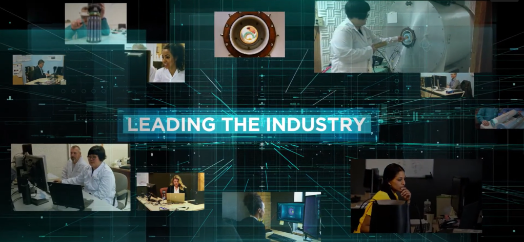
A Manitoba-based startup is making significant strides in medical imaging with the support of Mitacs interns. Co-founded by brothers James and Bob Schellenberg, Cubresa is advancing the field of brain imaging with the development of BrainPET — the world’s first clinical-stage positron emission tomography (PET) inserts for magnetic resonance imaging (MRI) systems.
From the early stages, innovative ideas contributed by Mitacs students played a crucial role in shaping the software methods that underpin BrainPET’s development.
One of those contributors was former Mitacs intern Sophia Krak, who helped calibrate the BrainPET scanner. The hands-on experience sparked a deeper commitment to neuroscience, leading her to conduct research on Parkinson’s-related dyskinesia and share her passion with youth through outreach.
“Thanks to Mitacs, I’ve been able to pursue my passion for neuroscience and medical imaging in both research and industry,” she says. “These experiences sharpened my technical skills, deepened my interest in brain imaging, and inspired me to give back.”
Designed for existing MRI machines, the technology’s portability opens up new possibilities for widespread use in various clinical settings, leading to enhanced accessibility and reduced costs.
Traditional MRI scans provide detailed images of anatomical structure changes that indicate advanced stages of disease. BrainPET, however, operates at the molecular level, detecting early biochemical changes before structural damage occurs. This capability is crucial for early diagnosis of conditions like Alzheimer’s, Parkinson’s, and epilepsy.
“An MRI shows you what already looks wrong — bones, tissues, organs — but that’s often when it’s too late. A PET scan detects changes at the cellular level before any visible signs appear,” explains James Schellenberg.
He adds, “Imagine rust forming on a car. The bubbling paint is what you’d see with an MRI. But PET shows you the molecules that cause the rust — while it’s still invisible to the naked eye.”

A personal connection to breast cancer drove James’s journey into medical imaging. In 2005, he filed his first patent related to breast cancer imaging and launched Cubresa. The company’s name reflects this mission: “Cu” for customized, “Bres” for breast, and “a” as a Latin feminine signifier. However, breaking into the breast imaging market proved to be a challenge due to dominating and well-established industry players.
After discussions with investors, Cubresa pivoted to brain imaging — a field with unmet needs and fewer barriers to entry. Since 2014, the company has been developing imaging solutions for neurological conditions, to improve early detection, diagnosis, and treatment monitoring.
Cubresa’s BrainPET technology was first installed at the Lawson Health Research Institute in London, Ontario. Initially a research-focused initiative, the project has evolved to have significant clinical potential.
Researchers at Lawson have been using BrainPET to image patients with epilepsy and brain cancer, gathering data that helps shape the future of what is possible for brain imaging. Each scan brings them one step closer to refining the technology and revealing new possibilities in diagnosis and treatment.
One of BrainPET’s most promising applications lies in epilepsy surgery. Surgeons seek to identify “foci” — areas of abnormal high brain activity — to target during procedures. While some foci are visible on traditional MRI scans, others only appear when PET imaging is incorporated.
By combining PET and MRI data, BrainPET provides a comprehensive map of brain activity, which is beneficial in precise surgical planning. Fusing two imaging modalities can significantly improve a surgeon’s ability to localize epileptic activity, improving surgical outcomes and patient quality of life.

Cubresa’s growth has been supported by long-term partnerships with academic institutions and initiatives like Mitacs. Over the years, the company has expanded,”reaching 20 employees in certain periods, and currently houses two former Mitacs interns.
“Mitacs has been important to us for over 10 years,” says James. “It allows us to jointly explore new challenges with a professor and test out emerging talent with fewer management demands and shared costs.”
These collaborations have allowed Cubresa to keep advancing its research and development efforts while helping shape the next generation of medical imaging talent. With innovation, partnership, and purpose guiding their work, Cubresa is opening a new chapter in the early detection of neurological disease — one that could redefine how we see the brain.
Mitacs’s programs receive funding from multiple partners across Canada. We thank the Government of Canada, the Government of Alberta, the Government of British Columbia, Research Manitoba, the Government of New Brunswick, the Government of Newfoundland and Labrador, the Government of Nova Scotia, the Government of Ontario, Innovation PEI, the Government of Quebec, the Government of Saskatchewan, and the Government of Yukon for supporting us to empower Canadian innovation.
Do you have a business challenge that could benefit from a research solution? If so, contact Mitacs today to discuss partnership opportunities: [email protected].
Mitacs’s website content is created by people throughout our organization, united in their passion for innovation and eager to share their perspectives with others in the innovation ecosystem.
Discover more stories about Mitacs — and the game-changing innovations driven by students and postdocs.


